
Scientific Lectures //
Optimizing and Standardizing Post-Mortem Magnetic Resonance (PMMR)
Natalie Adolphi, Ph.D. - Research Associate Professor, Department of Biochemistry & Molecular Biology, University of New Mexico
Presented: May 5, 2015
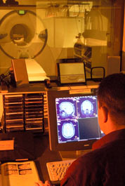
ABSTRACT: Magnetic resonance (MR) is notable for its excellent soft tissue contrast, and is therefore being investigated as a useful addition to the forensic imaging tool box. Examples of current forensic applications of PMMR include the imaging of fetuses and infants, anatomically complex soft tissues (e.g., brain and heart), and subtle soft tissue injuries that are not well-depicted by CT. However, it has been noted by a number of previous workers that MR tissue contrast obtained post-mortem (PM) often differs from that observed in MR images of live subjects obtained with comparable acquisition protocols, even when the PM tissue is not fixed. Therefore, efforts to understand and quantify the effect of normal post-mortem changes, such as reduced body temperature and decomposition, are ongoing. These basic and applied studies will enable PMMR acquisition and image processing protocols to move toward a higher level of optimization and standardization, which will facilitate systematic studies of the utility of PMMR for forensic investigation of specific types of cases.
After reviewing the current state of the art, we will present results from our ongoing MR study of unfixed PM brain tissue. In brain imaging, clinical T2-weighted imaging protocols yield good contrast between gray matter (GM) and white matter (WM) in the PM setting; however, when using clinical T1-weighted imaging protocols in the PM setting, poor contrast between GM and WM is obtained. The goals of this study include characterizing the temperature dependence of the tissue parameters T1 and T2, understanding whether temperature alone explains the poor T1 contrast between GM and WM, and adjusting PMMR acquisition sequences to improve endogenous T1 contrast. The temperature- and post-mortem interval (PMI)-dependence of the apparent diffusion coefficient (ADC) acquired from unfixed PM brain tissue will also be discussed.
To view presentation click here.

