COBRE Phase 1: MEGSIM //
MEGSIM
MEG is an excellent method for recording brain activity, with good spatial (mm) and excellent temporal (ms) resolution. However, the analysis of the MEG signal is complicated by the non-uniqueness of the electromagnetic inverse problem and by the fact that MEG systems manufactured by the three primary companies have different pickup coils, sensor arrays, noise cancellation methods, as well as different software packages for data analysis. Additionally, many MEG researchers either develop their own software for analyzing data or piece together shareware offered by others, partly due to the still unknown overall strengths and weaknesses of the different algorithms. This makes it difficult to relate results across laboratories and pool data together, particularly clinical data.
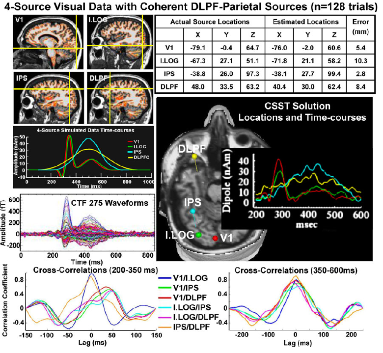
Simulation results for a 4-source model where all sources became synchronous during the later interval (see upper left panels for source locations (cross-hairs) and timecourses of the sources). Amplitudes and peak latencies were jittered across each of 128 single trials. The averaged waveforms seen at the sensor level for the CTF system are shown beneath the input timecourses. Upper right table shows CSST actual locations and errors associated with modeled source locations. The middle panel shows location and timecourse plots of the CSST solutions. Bottom row shows cross-correlations between source timecourses for an early interval (left) when there was some asynchrony across sources and a later interval (right) when all sources became synchronous.
The project (R21MH080141) aims to construct realistic simulated data sets in formats used by each of the 3 major MEG manufacturers. These can then be directly tested using various algorithms which include multidipole, spatiotemporal modeling, current reconstruction, minimum norm, and beamforming methods. The data sets and results are available to investigators worldwide via this download site, which will in addition act as a repository for results conducted by others for comparison.
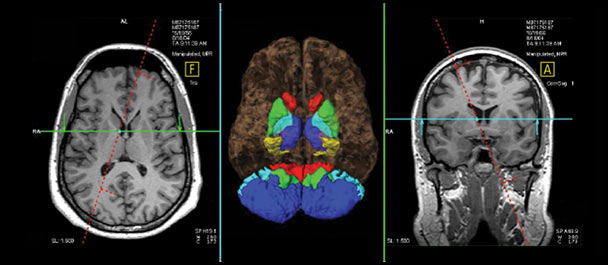
MEG and anatomical MRI data acquired from 5 subjects using basic visual, auditory and somatosensory paradigms are available via this site as well. The simulations and empirical data protocols were designed to activate either focal (4mm^2) or extended sources (20mm^2). The simulated signals are embedded in real MEG baseline recordings obtained from 5 healthy participants, using three MEG array configurations: VSM/CTF Omega (275 channels), Elekta Neuromag Vectorview (306 channels), and 4-D Neuroimaging Magnes 3600 WH (248 channels). Averaged data are available with zero noise and real spontaneous brain noise; single trial and continuous simulations are also available for a limited number of cases. The Mind Research Network (MRN) site will also be testing and posting the results of five different inverse procedures for comparison: multi-dipole spatio-temporal models (CSST), L1 minimum norm, two L2 minimum norm approaches sLORETA and SWARM, and a LCMV beamformer (Compumedics Neuroscan Curry 6.0).
Description of many of the simulations and results can be found here:
Evoked simulations and reconstruction results.
And our Neuroinformatics article can be found here:
Neuroinformatics article (2011)