Children’s Studies //
WELCOME TO OUR CHILD STUDY SITE WHICH DESCRIBES THE STUDIES WE ARE CONDUCTING TO BETTER UNDERSTAND BRAIN DEVELOPMENT IN YOUNG CHILDREN AND ADOLESCENTS.
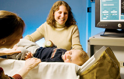
Infant Studies
Our research on children combines behavioral testing with safe and easy neuroimaging technology to better understand how the brains of infants, toddlers, and children develop. We are studying children with typical brain development as well as children with atypical brain development. Our studies currently focus on children diagnosed with Autism Spectrum Disorder, Epilepsy and Fetal Alcohol Spectrum Disorder. Our goal is to identify information that will lead to an earlier diagnosis of developmental disorders and to improve treatment options for young children diagnosed with epilepsy. We hope to improve the long term outcome for children affected by such disorders. As results become available we will provide updates on our current research projects. Please feel free to visit again for follow-up results on these studies.
 Child Studies
Our research on adolescents combines behavioral testing with safe and easy neuroimaging technology to better understand brain development individuals between 8 and 12 years old. We are studying normally developing individuals as well as those with atypical brain development. Our studies currently focus on children diagnosed Fetal Alcohol Spectrum Disorder. Our goal is to identify information that will lead to an earlier diagnosis of this disorder and lead to earlier, more effective treatments. We hope to improve the long term outcome for those affected by such disorders. As results become available we will provide updates on our current research projects. Please feel free to visit again for follow-up results on these studies.
Our research studies are approved by the Institutional Review Board at UNM Health Sciences Center. Participants are compensated for their time.
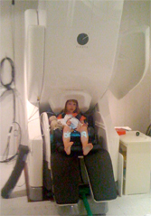
Why MEG?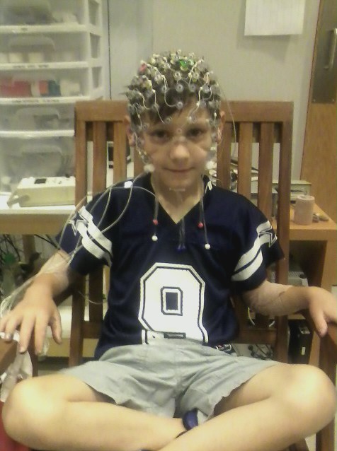
Magnetoencephalograpy (MEG) is a functional neuroimaging technique that allows us to collect data every 1/1000th of a second to monitor brain activity in real time. This technique, similar to electroencephalography (EEG) directly measures activity in the neurons in the brain. These techniques can be used to monitor resting brain activity or to examine the brain’s response to simple or complex stimuli (e.g. sounds, touch, visual stimuli). MEG is a much simpler compared to EEG because the child just needs to lie close to the sensors. EEG requires the attachment of electrodes/wires to the head. There is virtually no preparation time involved in MEG. MEG and EEG are both completely safe, non-invasive forms of neuroimaging.
 Technical Description
MEG is valuable in monitoring and investigating brain function because it is completely noninvasive, it has excellent temporal resolution (~1 ms) and good spatial resolution [0.5 cm; (Leahy et al., 1998; Stephen et al., 2003a)]. Since individual neurons transmit information on the same timescale, MEG allows one to monitor brain activity in real-time. EEG also provides fine temporal resolution, but MEG currently provides better spatial resolution of neural activity. In EEG, the skull and scalp blur the signal spatially because of the large changes in conductivity as the signal crosses these surfaces to reach the electrodes on the surface of the scalp. This is a particularly complex problem in young children and infants due to the anterior and posterior fontanels and sutures, which leads to additional distortion of the EEG signal (Flemming et al., 2005). The MEG signal, on the other hand, can pass through the skull and scalp virtually unaffected (Okada et al., 1999). Since the earliest age at which the sagittal and coronal sutures close completely is 6 and 11 years old, respectively (Hansman, 1966), EEG signals can change due to external factors through most of childhood. This variation in response due to differences in skull anatomy makes studies in young children difficult with EEG, whereas MEG results can be compared across different age ranges and participants without concern that differences in skull are generating differences in participant responses.